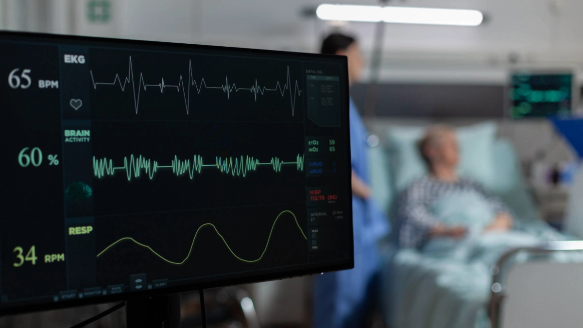
Echocardiogram in Turkey has become a widely trusted and accessible diagnostic procedure for evaluating heart health through high-resolution ultrasound imaging. At its core, this non-invasive test provides detailed insights into the structure, function, and motion of the heart and nearby blood vessels, helping physicians detect a wide range of cardiac conditions. In Turkish cardiology centers, especially at internationally accredited institutions, echocardiography is routinely performed using advanced equipment, handled by experienced cardiologists, cardiac physiologists, and certified sonographers. Whether a patient presents with chest pain, shortness of breath, or unexplained fatigue, an echocardiogram in Turkey ensures a comprehensive and real-time assessment of the heart’s performance, all without the use of harmful radiation.
What Is an Echocardiogram?
An echocardiogram—often called an “echo”—is a specialized ultrasound scan of the heart that utilizes high-frequency sound waves emitted from a handheld probe. As these sound waves bounce off the heart structures, they create echoes that are then translated into moving images, which are displayed in real time. Consequently, this non-invasive technique offers immediate insights into cardiac function. In particular, echocardiograms are indispensable in modern cardiology. In Turkey, they have become a foundational tool for cardiologists, as they enable detailed visualisation of heart valves, chambers, and blood flow dynamics. Therefore, echocardiograms play a critical role in both diagnosing and managing a wide range of heart conditions. Unlike an electrocardiogram (ECG), which records the heart’s electrical activity, an echocardiogram provides anatomical and functional imaging, helping doctors pinpoint structural abnormalities and determine the most appropriate treatment strategy.
When an Echocardiogram Is Used
An echocardiogram in Turkey is frequently used to diagnose and monitor:
- Damage caused by a previous heart attack
- Heart failure and reduced ejection fraction
- Congenital heart disease, including atrial septal defects and valve anomalies
- Heart valve dysfunction such as regurgitation or stenosis
- Cardiomyopathy (thickened or enlarged heart muscle)
- Endocarditis, a potentially serious infection of the heart lining
By visualizing how well the heart chambers contract and relax, and tracking blood flow through the valves and vessels, echocardiograms support early diagnosis and ongoing treatment decisions.
How an Echocardiogram Is Carried Out
Transthoracic Echocardiogram (TTE)
This is the most common form of echocardiogram. During a TTE, the patient lies on a bed while a sonographer applies a lubricating gel to the chest. A handheld ultrasound probe is moved across the chest to capture detailed images of the heart. Electrodes are attached to the chest to monitor rhythm throughout the test. The process is painless, takes 15–60 minutes, and typically requires no preparation. TTE is widely used across hospitals and cardiology clinics in Turkey due to its ease of use and reliable results.
Other Types of Echocardiogram
Transoesophageal Echocardiogram (TOE/TEE)
In cases requiring more detailed visualization, especially of the aortic valve or posterior heart structures, a TEE may be used. A thin probe is gently passed down the esophagus, close to the heart, providing high-resolution images. Patients receive local anesthesia and a sedative. While generally safe, this test may result in temporary throat soreness or, in rare cases, esophageal injury. Turkish hospitals follow stringent safety protocols during TEE procedures to minimize discomfort and complications.
Stress Echocardiogram
A stress echocardiogram combines imaging with physical or pharmacologic stress. Patients walk on a treadmill or receive medication to elevate the heart rate while echocardiographic images are recorded before and after exertion. This method reveals hidden heart disease, including ischemia and coronary artery blockages. It’s often used to assess how well the heart handles physical strain and can help determine treatment paths for chest pain or suspected coronary artery disease.
Contrast Echocardiogram
In a contrast echocardiogram, a harmless contrast agent is injected into the bloodstream. This agent enhances the visibility of heart chambers and wall motion, especially in patients with difficult acoustic windows. Turkish cardiology centres offer this advanced imaging service for more accurate diagnoses, particularly in obese patients or those with lung disease.
Fetal Echocardiogram
This non-invasive scan is typically performed between weeks 18–22 of pregnancy to examine the fetal heart. During the procedure, an ultrasound transducer is gently moved over the pregnant abdomen to capture detailed images. As a result, fetal echocardiography allows healthcare professionals to assess the structure and function of the developing heart. In Turkey, many advanced obstetric centers offer this service, combining high-resolution imaging with expert prenatal care. Consequently, fetal echocardiograms are instrumental in detecting congenital heart defects at an early stage, thereby enabling timely counselling, accurate diagnosis, and effective postnatal planning.
Echocardiogram Methods
Two-Dimensional (2D) and Three-Dimensional (3D) Echocardiography
Standard echocardiography starts with 2D imaging, showing the movement of the heart’s walls and valves. More advanced Turkish hospitals also offer 3D echocardiography, which reconstructs the heart’s structure from multiple angles. This is crucial before valve repair surgeries or assessing the function of the left ventricle.
Doppler Echocardiogram
This method analyzes blood flow and velocity using Doppler signals. It helps detect valve leaks, narrowing (stenosis), and pressure differences in heart chambers. Doppler echocardiography is a standard component of most echo exams in Turkey.
Color Flow Imaging
Adding a layer of visual clarity, color flow imaging reveals the direction and quality of blood flow through heart valves and chambers. It is invaluable for identifying regurgitation, turbulent flow, and congenital abnormalities.
Stress Echocardiography
In a stress echocardiogram, your heart is examined before and after exertion, either by treadmill exercise or with medications such as dobutamine. This test checks how well the heart copes with stress and helps uncover coronary artery disease, valve problems, or signs of early heart failure. Turkish cardiology departments closely monitor heart rhythm, blood pressure, and breathing during this exam, using contrast when necessary for improved imaging.
Three-Dimensional Echocardiography
3D echocardiography captures volumetric images, offering a more comprehensive view than traditional methods. It is commonly used for assessing congenital heart disease in children or planning complex valve surgeries. In Turkey, 3D echo is often paired with contrast or Doppler methods to enhance clinical interpretation.
Fetal Echocardiography
Fetal echocardiograms in Turkey are performed by maternal-fetal medicine specialists to detect congenital defects like ventricular septal defects or valve abnormalities. The procedure is entirely safe during pregnancy, and it allows for early intervention planning.
Risks
An echocardiogram is a safe, painless, and non-radiative procedure. The standard TTE poses no risks. However, minor side effects include skin discomfort from electrode removal or coldness from the gel. More complex procedures like TEE or stress echo have small associated risks:
- TEE: Sore throat, rare esophageal perforation
- Stress Echo: Dizziness, chest pain, irregular heartbeat
- Contrast Echo: Rare allergic reaction to contrast agent
Medical professionals in Turkish facilities monitor patients closely during these procedures to ensure safety and comfort.
Recovery After an Echocardiogram
In most cases, no recovery time is required. After a standard echocardiogram or stress test, patients can return to normal activities. For TEE, rest is recommended for the remainder of the day, and patients should not drive after sedation. Any throat discomfort typically resolves within a few hours.
Results
Once imaging is completed, the echocardiogram results are analyzed by a cardiologist. Results may be available within minutes or within a few days. Key findings include:
- Reduced heart function or wall motion abnormalities
- Valve dysfunction (stenosis or regurgitation)
- Abnormal chamber size or thickness
- Presence of blood clots or vegetations
- Pulmonary hypertension or restricted blood flow
- Congenital abnormalities
In Turkey, physicians use these findings to tailor treatment plans and may refer patients for further testing or intervention as needed.
Avicenna International Hospital – Excellence in Echocardiogram Services in Turkey
At Avicenna International Hospital, we are proud to be a leading provider of echocardiogram services in Turkey, combining state-of-the-art imaging equipment with world-class cardiology expertise. Whether you need a transthoracic echocardiogram, a detailed TEE, or fetal heart evaluation, our compassionate team ensures accuracy, comfort, and timely results. We deliver personalized cardiac care for both local and international patients, supporting early diagnosis, precise treatment, and long-term heart health. Avicenna is not just a hospital—it is a center of excellence for cardiovascular imaging and patient care.