Ophthalmology is an incredibly important field of medicine that focuses on the health and well-being of our eyes and visual system. Ophthalmologists, who are medical doctors specially trained in this field, are responsible for diagnosing and treating a wide range of eye conditions, including cataracts, hyperopia, glaucoma, macular degeneration, strabismus, and keratoconus.
To determine the overall health of a patient’s eyes, ophthalmologists conduct comprehensive eye examinations that assess visual acuity, eye alignment, and the structures of the eye. If necessary, ophthalmologists may prescribe glasses or contact lenses to correct refractive errors or perform procedures such as cataract surgery to remove clouded lenses. Lasers and intraocular lenses can also be used to correct a range of vision problems. But, treatments for glaucoma and macular degeneration can help prevent further vision loss.
Eye Examination
Comprehensive eye examination tests include the following:
- Visual acuity test
- Refraction assessment (measurement and evaluation of light refraction)
- Perimetry (microscopic examination of Visual field )
- Glaucoma screening (intraocular pressure)
- Retinal examination
What is a Cataract in Ophthalmology?
A cataract is an ophthalmology condition that occurs over the natural lens of the eye and makes the lens loses its transparency and makes it hard to see clearly. It is a clouding of the natural normal clear lens of the eye. it is common as people age. It is a bit like looking through a fogged-up or frosted window. Clouded vision caused by cataracts can make the vision status more difficult during reading or driving.
What are the symptoms of Cataracts?
The most prominent and common signs and symptoms of Cataracts are:
- Blurred or cloudy vision
- Double vision in a single eye
- Decreased vision
- Difficulty with vision at night
- Sensitivity to light or glare
- Frequent changes in the eyeglass degree or contact lens prescription
These ophthalmology symptoms usually appear according to age. Also, it can be seen in children, patients with diabetes, and patients who have sustained eye damage.
Cataracts treatment
It is performed either by using a laser, or eyeglasses or by cataract surgery, which is performed with the help of advanced techniques. In Femto Laser technology, which works with the help of computers and under microscopic magnification that enables the removal of the opaque lens by emulsifying it and the placement of a new, clear artificial lens in the eye with complete efficiency.
The most advanced technology is an important factor in the efficiency of the process and the satisfactory results for the patients’ lives. Moreover, Choosing an eye lens that replaces the natural lens that has lost its transparency and causes double or blurred vision. It is the most important factor in the efficiency of the process.
An intraocular lens (IOL) surgery can treat Cataracts and certainly, solves near- or far-sightedness problems in the same session. Thus, is divided into three categories: monofocal, bifocal and multifocal (trifocal). After cataract surgery, the patients will not have to wear eyeglasses again.
Laser and Smart Lens Treatments
Ophthalmology has played a vital role in the development and popularity of laser and smart lens treatments. Along with ageing, the natural lens in the eye loses its transparency and flexibility. it can no longer change shape or close focus or see near objects. It loses its transparency and develops Cataracts at later ages, usually after 40 years. The deformed natural intraocular lens after losing its elasticity can be replaced with Smart Lens.
This new multifocal lens will now be able to focus on different distances, the mid-distance and close. Thus both; the near and distant vision will be clear. The Smart lens, also makes the eye number never progress and prevents cataracts to develop in this eye. Ophthalmologists continue to explore new ways to improve vision through advancements in laser and smart lens treatments.
What is the Smart Lens?
Everyone has a natural lens inside the eye, which is both elastic and transparent, This natural lens focuses on distance sightedness due to this flexibility, thus can see both near and far views. With age over 40, the natural lens in our eyes first loses its flexibility, and cannot focus on close items and objects, as it loses its transparency and darkens gradually until vision loss occurs, and thus cataracts.
This problem occurs mostly over the age of 60 and beyond, and it may occur at early ages due to strokes or accidents in the eye, congenital causes, or inflammation inside the eye, and due to previous surgical processes. Importantly, the goal of the Smart lens procedure is to remove and replace the current deformed lens that has lost its function with an artificial one and gives the patient a better visual quality.
Who can have the Smart Lens?
In general, people over the age of 40 who have vision problems in the distance, near or both, and who would like to get rid of their eyeglasses for life, are the best candidates for this procedure. Anyone who has a Smart Lens will be able to see both distance and near on its own without the help of eyeglasses. In addition, for those who have undergone eye laser surgery previously, if they experience near or distant vision problems due to intraocular lens deformation, vision loss, or cataract problems, then the IOL is the first choice for them.
How is Smart Lens Surgery Performed?
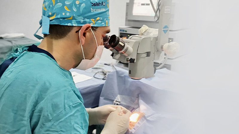
In intraocular lens surgeries, a very small microscopic surgical incision that does not exceed 3 mm is made to enter the eye and the membrane of the lens is removed in round form. The lens is inserted in the eye through the 3-mm incision and the body temperature transforms the lens into its normal shape.
The cataract surgeon enters the eye through the cornea to approach, and remove the cataract using ultrasound energy to emulsify the eye’s natural lens using the ultrasound waves of the Laser before placing the intraocular lens implant. Moreover, this new laser technology enables greater precision in creating the incisions required in traditional cataract surgery.
The laser, called femtosecond laser (FEMTO), is performed at the beginning of the procedure, it is painless and only lasts about 30 seconds. It provides more efficient cataract removal, requiring less energy and less stress on the eye, which results in a speedy recovery. The eyes are numbed with eye drops and the surgery takes about 10-20 minutes hospitalization is not required, and the patient is discharged on the same day after the procedure.
This modern cataract surgery allows the incisions to be tiny and makes it a completely painless operation. In which, the natural lens in the eye is extracted and replaced with a lens with multifocal functionality. Thus, both near and distant objects are placed clearly on the eye’s yellow spot, which provides visual acuity. Patients can adapt to the new lenses very fast and get rid of their glasses.
What is the importance of Smart lenses?
Provides a permanent solution and clear vision at near and mid, and far distances. The most important advantage of multifunctional smart lenses is providing distance clarity and can treat all types of Far-sightedness, astigmatism, hyperopia, myopia, or near-sightedness.
The most important characteristic of this corrective procedure is that it is suitable for all age groups, especially young people who should not have a FEMTO or LASIK procedure due to the incomplete growth of the cornea. Multifocal implanted lenses cannot be lost or replaced like eyeglasses or traditional contact lenses.
Cost of Intraocular Lens Operation
There are several things you should be aware of before conducting the price of IOL treatment, which are explained as follows: Our implantation materials and instruments that are used in our operations are all approved and certificated by the U.S. FDA agent and the smart lenses used during intraocular lens surgery are all imported from the USA and Europe. In addition, the devices used during surgery are imported from abroad. For this reason, the materials and devices consume more than 50% of the cost of the operations.
Some centres perform this surgery using lower-cost lenses, materials, and intraocular devices to provide a low cost for smart lens surgery. The materials that are produced in the Middle East or India are of low quality and low success rate which may lead to undesirable results in the future that cannot be remedied later. In our operations, we use the finest quality lenses that you will wear in your eyes for life. Smart lens prices vary according to the treatment to be applied depending on the condition of your eye. To get an accurate price, you must first visit our clinic and take an eye examination to fulfil your comfort and health.
Hyperopia
Ophthalmology has played a significant role in the development of advanced treatments for hyperopia, or far-sightedness, which is a common vision problem that often occurs with age. This condition happens when you can see distant objects clearly, but objects nearby and close to your eyes may be blurry. Far-sightedness usually presents at birth and resort to running in families. It can easily be corrected with contact lenses or eyeglasses. Another advanced treatment option is surgery. Visual disturbances due to ageing are no longer a problem thanks to surgical procedures performed with advanced technologies.
The “Hyperopia or presbyopia” surgery that can be performed from the age of 40 completely treats vision problems. Since it may not always be easily apparent that you are having vision trouble with your eyes, the ophthalmologist recommends intervals for regular eye exams, and If the degree of farsightedness is pronounced enough that you are not able to provide a task as well as you wish, or if the quality of vision decreases during your activities, the ophthalmologist will advise you of the options that are suitable to your case to correct your vision.
In the case of the surgery option, the lens placed in the eye with presbyopia surgery keeps the patient from wearing glasses for life. This process takes about 5-10 minutes, eliminates the possibility of developing cataracts in the future, and allows cataract patients to see good vision without glasses after surgery.
The lens in the eye is changing!
“Presbyopia” surgeries, are performed to eliminate problems with relative vision problems due to ageing and save patients from wearing glasses for life. In patients with cataracts, IOL is placed in the eye with the modern surgical technique termed Phacoemulsification (PHACO).
The patient’s lens is located behind the iris and is responsible for focusing light on the retina, producing clear, sharp images, and can change shape, once this lens deforms and loses its ability to accommodate and hardens, the option of PHACO surgery becomes the best solution. In this surgery, the lens is dissolved into particles and removed from the eyes, and an IOL is implanted and positioned in place. It is an outpatient surgery that does not need a hospital stay. In short, a new lens is positioned in place of the naturally deformed lens in the eye.
How does Presbyopia Occur?
The natural clear, soft, and flexible lenses that are easily changing shape and let you focus on objects both near and far away. The normal ageing process can cause presbyopia, especially after age 40, the lenses become more rigid and cannot change shape as easily as at younger ages. This makes it harder to do other near and close-up tasks like reading, driving, or threading a needle or any other applications and tasks that require near and close focusing.
What is the problem of farsightedness?
If Presbyopia or Hyperopia are not corrected, and the cornea or lens is not smoothly and evenly curved, the light rays will not be refracted properly, and you will have a refractive error which can lead to several serious problems after the age of 60 such as myopia (nearsightedness) and Astigmatism.
Frequently Asked Questions about Presbyopia
Are presbyopia and Farsightedness different?
The symptoms of presbyopia and farsightedness are similar, but these two conditions are not the same thing. Farsightedness occurs in people of any age, including babies, and children, it is when an irregularly-shaped eye (cornea or lens are not smoothly and evenly curved) prevents the light rays to be refracted properly and lined up with the retina. As a result, it’s hard to see items close up.
Presbyopia is an age-related problem in which the lens of the eye becomes rigid and less flexible. Adjusting focus between nearby and far-away objects and seeing details like reading words becomes difficult. Although, this problem is most common in people between the ages of 40 and 50. Presbyopia can be corrected with eyeglasses and contact lenses or refractive surgery
How Presbyopia is treated?
Presbyopia can be corrected with eyeglasses and contact lenses, eye drops, or refractive surgery. Difficulties in seeing close-up things appear and blurry vision start with age over 40, the easiest solution is to get a pair of eyeglasses that are used only for the near vision that is instructed by the ophthalmologist.
Thanks to advanced technology, the treatment of nearsightedness, hyperopia, and astigmatism with laser procedure can be performed within approximately 10 minutes. Again, a cataract can be removed with a short operation by the modern Phaco surgical technique.
Is Presbyopia Surgery difficult?
No, it is not a difficult surgery; the natural lens is removed in the eye to correct the eye number and replace it with a new lens. We prefer the surgery method for patients who are nearsighted or farsighted and whose eyes are not suitable for laser treatment. The process takes about 5-10 minutes and the patient begins to meet his relatives the next day.
Nevertheless, there are no cataract risks such as developing cataracts in the future since we remove the patient’s natural, faulty lens. Patients who suffer from vision problems can continue their lives now without wearing glasses.
Ophthalmology: Understanding Glaucoma and Its Treatment Options
What is Glaucoma?
Ophthalmology is the medical field that specializes in the diagnosis and treatment of eye conditions such as glaucoma. When the eye’s fluid builds up in the front part of the eye, which creates pressure inside the eye. As a result, this elevated pressure leads to damage to the optic nerve that is necessary for vision. If it is not treated early, it can lead to permanent loss of vision. Glaucoma disease occurs congenital or in adulthood.
Glaucoma Surgery
Once eye drops or oral medicine fails to lower the pressure inside your eyes, then surgery is an upcoming step. However, we apply various strategies of surgical and non-surgical techniques for treating glaucoma these include; (deep non-piercing excision of a portion of the sclera, trabeculectomy, minimally invasive glaucoma surgery, implantation methods, canaloplasty) with a success rate of more than 95% in Istanbul Hospital for Eye Diseases and Surgery Clinic.
Macular Degeneration
It is also known as “yellow-spot disease or age-related macular degeneration (AMD)” and is an eye degenerative disease that affects the central vision (reading area) of the retina (happens in the macula which is the central small portion of the retina) that affect the light-sensing nerve tissue at the back of the eye. It is very common over age 55 and can get worse over time. Thus AMD leads to severe and permanent vision loss if it is not treated early.
What are the causes of Macular Degeneration?
- Adults over age 55
- Genetics and a family history of AMD
- Smoking
Risk factors
The main risk factors for Macular Degeneration are:
- Ageing
- Genetic characteristics
- Hypertension
- Smoking
- A diet high in saturated fat
- High cholesterol
- Obesity
- Exposure to sunlight for long periods increases the incidence of the disease.
The factors related to age and genetics cannot be eliminated. However, other risk factors can be controlled. If there is high blood pressure, it can be controlled. If you smoke, quit. Sun-filtered sunglasses should be worn. It is recommended to avoid foods that contain butter, red meat, and cholesterol.
Symptoms of Macular Degeneration
- Visual distortions
- Vision distortion, straight lines may appear bent or wavy.
- Blurring of the central vision
- Poor vision quality
- Colour vision disturbances
What are the types of AMD?
There are two types of macular degeneration, the dry type, and the wet type.
Dry AMD is about 90% of age-related macular degeneration cases and the wet type is about 10%. However, early diagnosis of the wet type is more important because it can cause vision loss. Additionally, wet AMD progresses rapidly, and vision loss occurs faster than dry AMD. Furthermore, it can lead to blindness as a result of the accumulation of fluids and bleeding under the retina which can scar the macula.
How to treat AMD?
The macular degeneration treatment depends on determining the appropriate therapy for the patient, and this includes several therapy options such as:
Fundoscopy (or ophthalmoscopy) diagnoses and confirms the type of AMD and identifies the risk factors for vision loss. The surgeon may also require fluorescein angiography.
Early diagnosis of wet macular degeneration is of great importance because it causes vision loss in the later stages very quickly. The risk of developing AMD can be reduced by using vitamins that are rich in antioxidants and taking nutritional supplements such as lutein and zinc.
Also, Photodynamic therapy is a laser therapy combining injection of Verteporfin which is a photosensitive drug into the bloodstream. After that, laser treatment is applied to seal the leaky blood vessels and it is effective in treating wet AMD.
In addition, intraocular injections of some drugs such as Aflibercept injection can prevent leakage from newly generated blood vessels. The injections should be repeated 1 to 2 times per month.
What is a Fundus examination?
Fluorescein angiography includes the injection of a yellow dye (called fluorescein) into an arm vein, the dye travels through the blood vessels and a special camera takes the retina photos as the blood vessels took the dye colour.
By passing a light beam that provides a complete view of the entire retina. This shows if abnormal or new blood vessels are growing under the retina. However, it confirms an early diagnosis and provides early treatment to reduce or slow the incidence of AMD.
The Amsler Grid Test in Macular degeneration
It is a simple tool used for the detection of vision problems resulting from macula damage to detect early signs of AMD or any other eye disease. However, It is an easy test that you can apply at home. Importantly, adults over the age of 40 should do this test.
Amsler Grid Application
- Wear eyeglasses or contact lenses that you usually use and wear for reading.
- Hold and keep the Amsler grid approximately 14 to 16 inches from your face and under normal room lighting.
- Close one eye with your hand and focus on the dot in the centre of the grid.
- Keep your eye focused on whether you can see the four corners of the large square are clear
- Test your other eye the same way.
- Irregularities and Blurry lines.
- If you can’t see the corners, it is essential to consult an ophthalmologist as soon as possible.
Strabismus: A Condition Treated by Ophthalmology
Strabismus or crossed eyes is when one eye is turned in a direction that is different from the other eye. Patients with strabismus have problems with controlling eye movement and are not able to keep normal eye positions. However, the Strabismus can be classified by the direction of the misaligned or turned eye including; Upward turning, Downward turning, Inward turning and Outward turning.
Moreover, It usually appears in young children and infants, and most often in children who are 3 years old, and even adults can develop strabismus. On the other hand, the treatment options for strabismus can include different methods including using special contact lenses or eyeglasses, medications such as eye drops, injections, or ointments, or eye muscle surgery that changes the position or length of eye muscles and makes them aligned correctly.
However, this surgery was performed under general anaesthesia. it is also possible with local anaesthesia. Therefore, surgeries do not require a long hospital stay, and the healing process will be fast. Patients return to their normal activities within a few days.
Glasses may be required after surgery. In some cases, a second surgery may be required at a later time. Early surgery gives better results for children with fixed squints. However, eye muscle surgery may have some risks.
Keratoconus: A Condition Treated by Ophthalmology
The anterior region (front surface) of the eye consists of a clear, transparent, dome-like layer called the cornea. This transparent layer of the eye helps to bend and focus light rays onto the retina. Any problem or abnormality in its shape leads to visual issues. Visual functions occur with the intact cornea layer. In other words, the cornea thins, and its dome shape starts to bulge and become cone-like instead.
This process of re-shaping and thinning causes sensitivity and glare to light, this condition is called Keratoconus. Keratoconus is a degenerative, progressive disease that usually begins at the age of 20 and develops. Consequently, it causes focusing and vision problems.
Moreover, the transformation of the convex shape of the cornea to a conical shape leads to an increase in myopia, astigmatism, and distorted and blurred vision problems, which progress unless treated.
What are the required tests before Keratoconus surgery?
The ophthalmologist may require the patient’s medical history at first. Then performs an eye examination to diagnose and decide on an appropriate treatment for keratoconus.
Therefore, the surgery and treatment methods are planned according to the patient’s condition. There will be several tests that your doctor may require to diagnose the state of the cornea. these Include:
- Keratometer (to measure the degree of the cornea curvature)
- Corneal topography
- Fundus examination
- Slit Lamp examination
- Visibility level without glasses
- Eye refraction test (measurement of the refractive error in the eye with and without drops)
How is Keratoconus treated?
Improving the vision depends on the keratoconus severity. In the early stages of keratoconus, it can be treated with only eyeglasses or contact lenses to correct vision problems.
Unfortunately, this is a progressive condition and in some people, vision will eventually worsen, and the cornea becomes scarred rapidly. In these situations, cornea transplant surgery might be necessary.
Since keratoconus is a progressive disease, an early diagnosis is very important and depends on the previously mentioned examinations. Especially the topographic examination of the cornea. In addition, the necessary keratoconus operations are performed to stop or slow the progression of the disease.
Treatment Methods of keratoconus.
Corneal collagen cross-linking
the cornea is saturated with a combination of riboflavin eye drops (vitamin B2) and collagen that are treated and activated with ultraviolet light. Therefore, causes cross-linking of the cornea significantly and safely strengthens the rigidity and stiffens the cornea to prevent further shape changes.
Consequently, helps to decrease the risk of progressive vision loss via stabilizing the cornea which helps to stop the keratoconus progression. It is an outpatient procedure performed in the clinic under eye drop anaesthesia. Patients usually experience some mild discomfort, it takes approximately one hour.
Corneal transplantation (keratoplasty)
Surgical options are decided depending on the severity of the eye condition and the location of the puffy cone. It involves the clear plastic segments insertion into the cornea which is fixed by a small incision. However, these segments are designed and made to reshape the front surface of the eye. Therefore, correcting refractive errors that happened due to keratoconus. Most importantly, it is used for advanced cases of keratoconus, where the patient’s vision is not correctable with contact lenses or glasses.
Today a FEMTO Laser is applied and the incision is created within approximately 10 seconds and the surgery is performed within 5-10 minutes. Thus, there is no need for stitches and very little pain occurs. The main purpose of treating the corneal ring in patients with advanced keratoconus is to increase the patient’s visual quality
After keratoconus surgery
Post-keratoconus surgery, the necessary instructed medicines must be used regularly. Since the surgical keratoconus intervention is performed on the eye, rubbing the eye should be avoided. Therefore, avoid sun exposure and microbial environments as much as possible to prevent the dangerous situations that may occur after keratoconus surgery.


