Cardiovascular Medicine
The Cardiology Department at Avicenna International Hospital provides all the diagnosis and treatment services for cardiovascular diseases. Avicenna hospital provides you with everything related to cardiovascular medicine and surgery with the most modern medical technologies.
Outpatient Clinic
Avicenna’s Cardiology unit provides the diagnosis and treatment of a wide range of cardiac muscle problems and diseases by the most experienced specialists. Examinations are performed by our specialists and physicians six days a week (except Sunday) from 09:00 to 17:30.
Electrocardiography (ECG)
It is an examination of the electrical activities of the cardiac muscle to diagnose its performance and the neural conductivity system. ECG is an important method of diagnosis of various conditions of the heart and cardiovascular diseases, for instance; rhythm disruption, heart failure, heart valve diseases, and cardiovascular occlusion
Stress Test
It also knows as the “Stress ECG Test”, “Exercise Test” or “Treadmill Exercise Stress Test”. It helps to investigate and diagnose the existence of cardiovascular problems and diseases and determines the most effective treatment method according to the patient’s cardiovascular situation, for instance;
- Irregular heartbeat
- Disturbances in the heart muscle
- Patients suffering from high blood pressure
- Heart valve diseases.
- It also, detects whether arrhythmia happens due to stress and physical activity
- Detects any disturbances of the blood flow within the heart muscle.
During the stress test, the patient performs exercises such as walking on a treadmill or riding a stationary bike, as the exercise speed is adjusted by the physician to elevate the heart rate. Thus, it enables the identification of abnormal detections that cannot be identified in ECG at rest, and after the exercise.
The exercise test provides a great detection in the early diagnosis of cardiac diseases. The results are given immediately after the examination.
How to Prepare Before the Stress Test Procedure?
- Do not eat, drink or smoke at least 3-4 hours before the stress test.
- Bring your previous stress test results, if you had any.
- Take all kinds of the prescribed medications you have been using previously with you to the hospital. Ask your doctor whether it is safe to continue taking them.
- Wear comfortable clothes and walking shoes.
Echocardiography (ECO)
Echocardiography is an examination of the heart’s structure and performance through the ultrasound waves that are traveling to the heart and are checked via the echocardiography device. However, the device uses ultrasound waves to produce images of the heart, and it enables the doctor to have information about the
- cardiac vessels
- heart muscle
- cardiac valves and to identify any disorder among them.
There are several types of echocardiography which will be mentioned as follows:
Transthoracic Echocardiography (TTE)
During this method of echocardiography examination, the patient is required to sleep on a stretcher. Then, the water-based gel is spread on the transducer device by the technician. However, the technician applies the device firmly on the chest area in different positions to examine the heart with very detailed information by recording the echoes of the heart.
Nevertheless, it is a safe method and does not involve any radiation, and it can be performed on anyone, involving pregnant women or newborn babies. It does not include any pain or side effects. Also, there is no medication used during the process and does not require any special preparation.
Echocardiography is performed at the Avicenna International Hospital using the most advanced echocardiogram devices in the world to diagnose heart structural diseases, heart failure, congenital heart disorders, aorta problems, and heart valve diseases. The result is obtained immediately after the examination.
Stress Echocardiography
It is another type of echocardiogram test used to investigate and check whether there is a blockage or constriction in the heart vessels and arteries feeding the cardiac muscle.
However, it is performed through physical activity by simply diagnosing the heart with ultrasound waves before and after the stress that is made by walking on a treadmill.
Thus, the stress is made through exercising on a treadmill device or applying some drugs to increase the work burden on the heart muscle.
It is used to examine whether other treatments might be required other than drugs in patients with myocardial infarction, and to identify the severity of the heart’s problem in valvular heart diseases. The result is obtained immediately after the examination.
How to Prepare Before Stress Echocardiography?
- Do not eat, drink or smoke before the stress test.
- Consult your doctor about all the prescribed medications you have been using.
- Wear comfortable clothes and walking shoes.
- Bring the results of your previous heart tests
Transesophageal Echocardiography (TEE)
TEE echocardiography is used when the physician is requiring more accurate and clearer images of the heart that is can not be visualized with TTE or ECG due to the structure of the chest. However, to examine the intra-heart formation clearly and closely.
Therefore, this type provides better visualization of the heart chambers and gives a more detailed echocardiographic estimation that is made from the oesophagus.
Initially, local anaesthesia is made by spraying the mouth to relieve pain, then, a thin tube containing the endoscope is inserted into the mouth through the oesophagus until it reaches the heart’s level, and clear and very detailed 3D images of the heart are obtained.
Moreover, it takes 30 minutes including the preparation before the test. The result is given immediately after the examination.
How to Prepare Before the TEE test?
- Do not eat, drink or smoke at least 3-4 hours before the stress test.
- If there is a prosthesis in your mouth, remove it before the examination.
- Consult the doctor if you have swallowing disorders or diabetes problems.
- Bring the results of your previous heart tests.
How to Prepare After the TEE test?
- Do not eat for two hours after the examination
- Do not drive for a few hours until the effect of the anaesthetic medication wears off
The 3D transesophageal echocardiography method is only available in very few health centers in our country, including Avicenna Hospital / Atasehir, as this method provides detection of congenital heart abnormalities, leaks in artificial heart valves, disorders, and problems in the heart valves.
Holter Monitor
It is a small device that is worn for one or two days according to the time that the doctor requires to record the heartbeats and monitor the heart rhythm. Thus, it keeps tracking the heart rhythm while the patients continue their daily life and usual activities.
Sometimes; smartwatch devices can offer electrocardiogram monitoring. However, the doctor uses the captured data on the Holter monitor to find out if you have a heart rhythm disorder.
It is a non-invasive and painless method. It involves using 4-6 electrodes (sticky patches) that are attached to the chest and sense the heartbeat. These electrodes are connected to a recording Holter device with wires (the wires are made of soft plastic 3-4 cm in diameter).
While the person resumes his/her normal daily life activities, the device particularly records all the heart data and saves it for later reading by the physician after the specified period expires. Finally, when the specified period ends, the device is removed by the doctor.
Thus, information and data recorded by the Holter device are analyzed on the computer to see if there is a problem with the heart rhythm.
However, the device can detect heart problem symptoms, skipped heartbeat, almost all kinds of arrhythmias, irregular heart rhythm, and other problems such as palpitations, chest pain, and fainting. Moreover, this device can detect all the heart irregularities that electrocardiography couldn’t identify or detect.
Blood pressure Holter
it enables hypertension diagnosis and treatment efficiency via recording the blood pressure and pulse during daily activities, sleep, and during rest at regular intervals for 24-72 hours throughout the day. Therefore, it is determined at any time of the day. It helps in providing early diagnosis and setting a treatment plan.
Tilted Table Test
It is a test performed to investigate the reasons behind fainting caused by a sudden change in blood pressure and arrhythmia after prolonged standing or sitting. The tilted table test is performed by lying on a table that can be tilted (moves in an upright position) and placing electrodes on the chest and arms.
However, excessively low blood pressure and/or pulse rate indicate an abnormal response to cardiovascular disease.
Pacemakers
Lots of people around the world have pacemaker implantation. These small and high-tech devices are used for many purposes, for instance; preventing arrhythmia, treating myocardial infarction and heart failure, acting as a pump, and prohibiting sudden and unknown-reason deaths.
Moreover, it helps the patients to return to their regular life by elevating the life quality. There are 3 types of pacemakers:
- Single chamber: to eliminate slow heart rate
- Double Chamber Pacemaker: for treating heart failure
- Implantable cardioverter Defibrillators (ICD) are used in arrhythmia cases. It gives electroshock when the heart fails to perform its function.
A pacemaker is implanted in patients with heart arrhythmia who cannot administer their lives normally. However, people with pacemakers can resume their normal lives such as; household chores, going to work, swimming, travelling, driving, and having sex lives.
Pacemaker Information
While travelling, patients should discover the nearest clinics in their destination in case any unfavourable conditions happen, because pacemaker performance must be monitored after it is inserted.
The pacemaker, which is a small computer, can be read and monitored from the outside with the assessment of another computer using a telemetric method. Thus, information such as the patient’s heart rate progression, if there were any other rhythm disorders, does the patient have arrhythmia from time to time, the pacemaker working period, or was the patient was always connected to the pacemaker.
Besides, it is possible to program the pacemaker on the appropriate values to keep the rhythm regular and on the required volts of the battery. All models of the pacemakers batteries can be found and monitored at Avicenna International Hospital.
Catheter Ablation
The method of supraventricular tachycardia ablation is used in the treatment of rhythm disturbances that cannot be controlled with drugs or in some cases when patients refuse to take medication for life.
However, it is used to treat arrhythmia and rhythm disorders through radio waves. The energy of the radio-frequency waves forms small scars within the heart to obstruct irregular or abnormal electrical signals and retrieve a normal heartbeat.
Moreover, when the arrhythmia is so serious that it can threaten the patient’s life, a direct catheter method might be required. The method is primarily involving the numbness of the needle insertion points via local anaesthesia, yet in some cases, under general anaesthesia. Moreover, during the operation, sedative drugs may be used.
The success rate for treating arrhythmias problems by applying this method is between 70-100%. After a successful application, the probability of the recurrence of arrhythmia which depends on the arrhythmia type ranges between 3-5%.
At Avicenna International Hospital, in addition to the radio waves ablation method, other treatments of arrhythmias can be performed using the “cryotherapy” method called cryoablation, coronary balloon catheterization, or angioplasty.
Acute rheumatic fever disease
Rheumatic fever is an inflammatory immune disease that mostly affects school-age children between 4-14 years. Rheumatic fever begins with inflammation in the throat and pharynx and causes damage to the heart valves. It includes stenosis a way that makes blood passage difficult through the heart’s chambers.
Therefore, it results in heart symptoms in the future as a result of injury of the heart valves and leads to fluid accumulation around the heart. However, results in chest pain, tachycardia, and shortness of breath. Sometimes drug treatment is not sufficient for chronic cases, so Mitral Balloon Valvuloplasty or open-heart surgery is performed.
Valvuloplasty or angioplasty is an invasive surgical procedure performed by inserting a catheter through the groin (thigh) or arms to expand the narrowing of the artery through a catheter with a special balloon that is sent to the narrowed artery and the balloon is inflated to expand and open the tightened valve. The wire movements are monitored on the computer screen. Balloon therapy results are successful and the patients will feel much better.
Advantages of Catheter Ablation
- It is performed with local anaesthesia and the patient remains unconscious during the procedure.
- A small incision (hole) is made in the right or left thigh, and the balloon is pushed into the heart through this hole
- There is no need to perform risky open-heart surgeries and open the thoracic rib cage.
- After the operation, patients are kept under observation.
- Patients can stand the next day of the operation.
- The majority of patients can be discharged the following day.
- There is no need to use blood thinners after the procedure.
- With balloon valvuloplasty alone, 90% of patients decline complaints. This improvement can last for up to 20 years. Most patients feel comfortable for at least 5 to 10 years.
Coronary Angiogram
It is a method used to detect cardiac vascular disorders through X-ray imaging. Coronary angiography diagnoses the obstruction and narrowing of the arteries that feed the cardiac muscle. whether an artery is narrowed or blocked.
However, this test is a part of the group of procedures named heart catheterizations. Cardiac catheterization can both treat and diagnose blood and cardiac vessel conditions.
A coronary angiogram is the most common type of heart catheterization which diagnoses heart conditions and treats heart problems.
In this imaging, a coloured material (dye) is injected, which appears in the X-ray imaging, and images are taken of the blood vessels, which allows the vessels to be seen.
During a coronary angiogram, the patient lies on the X-ray table, and a type of dye that can be visualized by an X-ray machine is injected into the blood vessels of the heart. The X-rays take a series of pictures from several angles providing an accurate look at the blood vessels.
Moreover, the intervention area is numbed with local anaesthesia and a small incision is made to insert the catheter (a short plastic tube) into the artery of the arm to enter the blood vessels and coronary arteries. Then the dye is injected through this catheter so that the blood vessels can be easily seen on the X-ray images.
However, the coronary angiogram is completed after 30 to 60 minutes, the patient is transferred to the recovery room for observation and the patient hospitalization is a must for this test. The patient should rest for 4-6 hours after the examination, and it may take longer, for example; for those who have undergone various previous heart surgeries.
Percutaneous Transluminal Coronary Angioplasty (PTCA) and Stent
It is a procedure used for treating the narrowing or blocked cardiac vessels after its detection through a coronary angiography test. PTCA is performed under local anaesthesia and by using special materials.
First, the physician makes an incision through the arms, wrist, or groin areas and inserts a balloon catheter through the intervention site into the blocked vessel to widen the vessel. However, The doctor will inject a dye through the catheter to see the inside structures of the blood vessels and detect the stenosis with angiography.
Later, the balloon is inflated to widen the vessel, and therefore, the stenosis or clogging disappears. Moreover, usually, people how undergoing angioplasty are also having the placement with a very thin and tiny mesh wire called the Stent, which is collapsed around the balloon. While the balloon expands inside the blood vessel, the stent expands and is placed within the artery, therefore, the blockage will disappear.
In the past, the stents used were made of stainless metal only, but now, according to technological development, new and distinct stents that are soluble in quality and involve drugs are used. The physician decides the type of stent according to the patient’s needs and condition.
PTCA is an application that involves hospitalization for one to two days. The patient is discharged once the doctor sees it is appropriate. Patients need to avoid stressful environments such as exercising, working, and sexual intercourse for 15 days. The doctor determines the date when the patient can travel by plane.
Transcatheter Aortic Valve Replacement (TAVR)
Also known as Transcatheter Aortic Valve Implantation (TAVI). It describes the treatment of aortic valve stenosis or the restricted aortic valve by a minimally invasive heart operation of replacing or implanting the aortic valve in the heart using the catheterization method avoiding the high risk of open-heart surgery.
However, in this method, a small incision is made in the groin or through the chest which is 4-5 cm in diameter under local anaesthesia and they will give the patient an IV medication to eliminate blood clotting. Then, the surgeon will access the heart through the intervention point and insert a catheter until it reaches the aortic valve of the heart.
However, the catheter movement inside the heart will be guided and monitored with the help of advanced imaging techniques. When the new valve is placed, the balloon of the catheter will be inflated to expand the newly implanted valve into its correct position. As a result, the catheter is removed when the surgeon is sure about the valve position.
After the procedure
the patient might spend the night for monitoring purposes in the intensive care unit. Moreover, Prescribing some blood thinners to prevent blood clots, and the patient should regularly come to the hospital for follow-up appointments with the doctor a few days after TAVI.
The TAVR method is recommended for patients who are at high risk for open-heart surgery, for example; elderly patients, patients with lung, liver, or kidney disease, or patients who have previously undergone open-heart surgery.
In our Cardiovascular department at Avicenna International Hospital, the determination of treating aortic stenosis is made after the consultation with a heart and heart surgery specialists team, to find out the best treatment option for the patient and to alter the signs and symptoms of aortic valve stenosis to improve people lives quality.
Non-Surgical Methods of Vascular Disorders
Today, the most preferred treatment methods are non-surgical procedures to avoid high-risk open-heart surgeries, especially for those at high risk for open-heart surgery. Non-surgical treatment methods are mainly suitable for patients with congenital holes within the heart.
Also, conditions such as an internal aneurysm or ruptured blood vessels require urgent surgical intervention. However, the surgery option cannot be applied to every patient. Because the patient’s vascular structure must be suitable for surgical treatment.
Therefore, minimally invasive reconstructive procedures are performed that can be done under local anaesthesia in a sterile hospital environment and operating room conditions. There are two techniques for vascular treatments: Endovascular Aneurysm Repair (EVAR), and Thoracic Endovascular Aortic Repair (TIVAR)
The option of TEVAR requires an incision through the chest or breastbone or groin. The options are applied depending on the type of disease and usually take around 2 hours to complete.
At Avicenna Hospital, the holes are closed with the help of the most modern medical devices with a catheter.
This treatment method has a shorter recovery time than open-thoracic surgery, the patient is discharged within 24-48 hours after the procedure. After recovery, patients should regularly come to the hospital for follow-up appointments.
Renal Sympathetic Denervation (RSD)
Renal denervation is a minimally invasive intervention in patients who have arterial hypertension. There are nerves called “sympathetic” that cause elevated blood pressure around the renal vessels.
Therefore, this method relies on burning these nerves in the renal artery, which causes high blood pressure, without causing any damage to the arteries themselves using special materials and x-rays. However, this procedure is effective in persistent hypertension, which critically affects a patient’s life quality
We provide an 80-90% success rate in treating and controlling stubborn blood pressure and reducing the number of blood pressure drugs and medications used by patients.
Nuclear Medicine Department
Nuclear Stress Test (Thallium Test)
Department of Nuclear Medicine at Avicenna International Hospital provides a better diagnosis of the risk of a heart attack, coronary artery disease, or other cardiac problems, and guides the treatment of these disorders and diseases if the patient has been diagnosed with any. However, the doctor may require the nuclear stress test from the patient once a routine stress test could not indicate the reason behind several symptoms such as breath shortness or chest pain.
It includes the use of a radioactive dye and an imaging machine to produce images displaying the blood flow to the heart. Therefore, it measures the blood flow toward the heart via two sets:
- The first set of imaging is while the heart is at rest, and the other set is after stress (overwork) through exercising. Nevertheless, to show any damage in the heart and also, the poor blood flow areas.
Usually, it involves the insertion of an intravenous injection of the radioactive dye into the arm, which may feel cold at first. However, it is absorbed by the cardiomyocytes after 20 to 40 minutes. Then, the technician will put electrodes on the chest, arms, and legs while the patient is lying still on a table to start taking the first imaging set of the heart (when the cardiac muscle is at rest).
- The second imaging set is done when the heart exerts effort while walking or running on a treadmill or a stationary bike. After completing the test, the doctor will advise you to drink large quantities of water to excrete the radioactive dye from your body through urination.
- However, if the rate of blood flow to the heart is insufficient; You will undergo coronary angiography. If there are severe obstructions, then there may be a need for surgical intervention (open chest surgery).
- By-pass
- Heart Surgery
- Valve Surgery
- Aort Surgery
- Carotis Artery Surgery
- Peripheral Artery Surgery
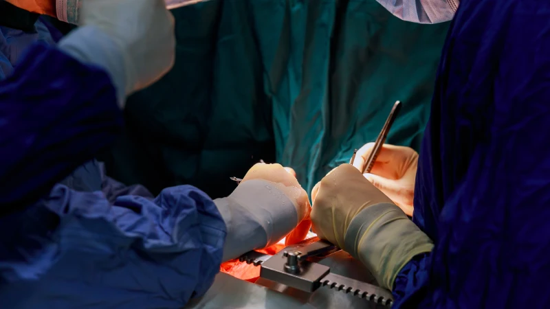
We open the roads to your heart….. Avicenna Hospital, is at your service with the latest technology in cardiology. Our team of experts are proud in contributing their knowledge and skills towad risk reduction invasive procedures.CARDIOVASCULAR SURGERY We open the roads to your heart….. Avicenna Hospital, is at your service with the latest technology in cardiology. Our team of experts are proud in contributing their knowledge and skills towad risk reduction invasive procedures.
CARDIOLOGY
Effort tests are carried out in cases of suspected artery constrictions. Echocardiographic ultrasound and doppler analysis are carried out to establish the conditions of heart-related dysfunctions. Arythmic disorders, associated with hypertension are evaluated with Holter monitoring. NONINVASIVE CARDIOLOGY UNIT Diagnosis and therapy
- Cardiology Clinic
- Effort test lab
- Echocardiograpy lab , Colored Doppler Echocardiography, Contrast and Stress Echocardiography
Contrast and Stress Echocardiography,
- Holter Monitors. Rythm Holter, Blood pressure Holter
Therapeutic Procedures
- Coronary Angioplasty
- Angioplasty-stent implentation.
- Temporary and permanent pace- maker Implants
- Coronary intensive care unit
- Primary PTCA+Stent
- Pulmonary baloon valvuloplasty
INVASIVE CARDIOLOGY UNIT Diognosis and therapy
-
- Coronary Angiography
- Heart Catheter
- Perypheral angiography




Leave a Reply