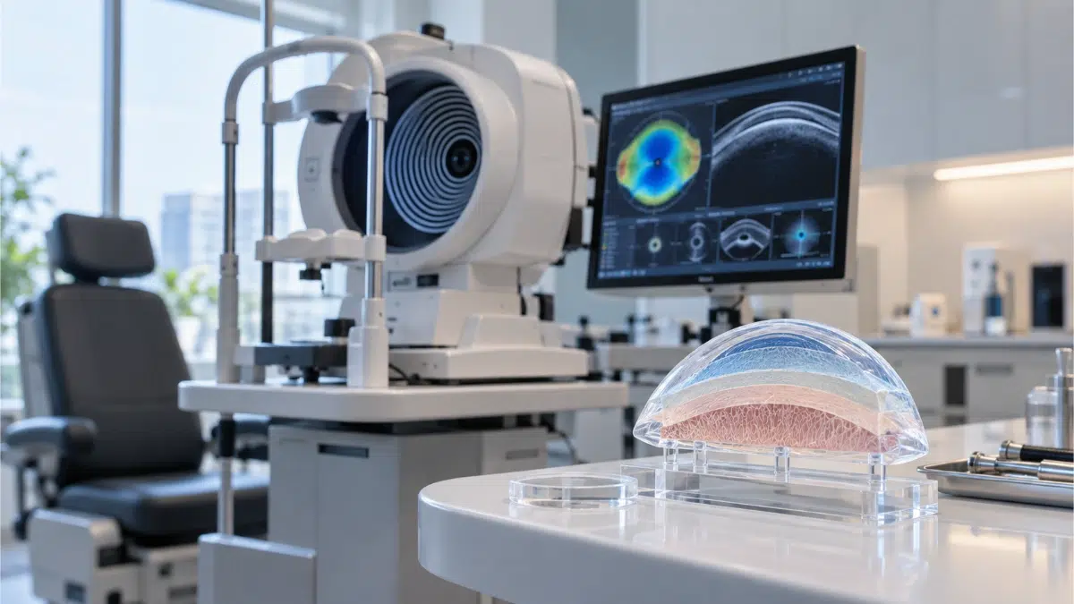


Laser eye surgery is planned from more than a glasses prescription. Before LASIK, PRK or another surface-laser procedure is considered, the ophthalmologist needs to understand the shape and structural characteristics of the cornea.
Corneal topography creates a detailed map of corneal curvature. Corneal tomography provides additional three-dimensional information, including the front and back surfaces of the cornea and how corneal thickness is distributed.
These measurements can reveal abnormalities that may not be obvious during a routine eye examination. They are particularly relevant when screening for keratoconus, irregular astigmatism, contact-lens-related distortion and other corneal patterns that may affect whether refractive surgery is appropriate.
Corneal Topography Before Laser Eye Surgery: Quick Answer
Corneal topography maps the curvature of the front surface of the cornea, while corneal tomography provides additional three-dimensional information about the front and back surfaces and the distribution of corneal thickness. These tests help identify abnormal corneal patterns before LASIK, PRK and other refractive procedures. Corneal thickness alone is not enough to determine whether laser eye surgery is appropriate.
What Is Corneal Topography?
Corneal topography is a non-invasive imaging examination that maps the curvature of the corneal surface.
The cornea is the transparent front part of the eye and contributes substantially to its focusing power. Small differences in corneal shape can therefore affect how light is focused.
Topography systems measure multiple points across the corneal surface and convert them into maps that help the ophthalmologist assess:
- Corneal curvature
- Relatively steep or flat areas
- Corneal symmetry
- Regular and irregular astigmatism
- Patterns that may suggest keratoconus or another abnormality
Topography is commonly used during assessment for refractive surgery.
What Is Corneal Tomography?
Corneal tomography evaluates the cornea in greater structural depth.
Traditional topography primarily describes the curvature of the anterior, or front, corneal surface. Tomography can provide information about:
- The anterior corneal surface
- The posterior corneal surface
- Corneal elevation
- Thickness across different areas
- Location of the thinnest point
- Overall three-dimensional corneal shape
Depending on the system, tomography may use technologies such as Scheimpflug imaging, scanning-slit imaging or anterior-segment OCT.
This additional structural information can be particularly useful when assessing whether the cornea appears stable enough for refractive surgery.
Corneal Topography vs Tomography: What Is the Difference?
| Feature | Corneal Topography | Corneal Tomography |
|---|---|---|
| Main information | Surface curvature | Three-dimensional corneal structure |
| Anterior corneal surface | Yes | Yes |
| Posterior corneal surface | Limited or not directly measured by traditional systems | Yes |
| Thickness distribution | Limited depending on system | Yes |
| Thinnest-point assessment | Not the main purpose | Commonly included |
| Keratoconus screening | Useful | Adds structural information |
| Refractive-surgery planning | Important | Often complementary |
The two tests are not simply competing versions of the same measurement.
An ophthalmologist may use information from several imaging tests together when deciding whether laser eye surgery is appropriate. Pasted text
Why Is Corneal Imaging Important Before Laser Eye Surgery?
Laser eye surgery reshapes corneal tissue.
Before altering the cornea, the ophthalmologist needs to determine whether its existing shape and structure appear suitable for treatment.
Corneal imaging may help identify:
- Keratoconus
- Early or subtle ectatic patterns
- Irregular astigmatism
- Contact-lens-related corneal warpage
- Other abnormal corneal shapes
Abnormal topography or tomography can be an important finding when assessing the risk of corneal instability after refractive surgery.
This is why treatment should not be selected from prescription numbers alone.
Can a Routine Eye Examination Miss Corneal Abnormalities?
Yes.
An eye may appear relatively normal during a routine examination while corneal imaging reveals a suspicious pattern.
This is particularly relevant in early or subclinical keratoconus.
Structural changes can begin before obvious abnormalities are visible during a routine slit-lamp examination. Topography and tomography provide additional information about curvature, elevation and thickness distribution.
However, an unusual map does not automatically mean that keratoconus is present. The findings need to be interpreted together with the rest of the eye examination.
What Is Keratoconus?
Keratoconus is a condition in which the cornea progressively thins and changes shape.
Instead of maintaining its usual curved profile, part of the cornea can become increasingly steep or cone-like.
Possible effects include:
- Increasing myopia
- Irregular astigmatism
- Distorted vision
- Reduced visual quality
- Difficulty achieving clear vision with ordinary glasses
Corneal imaging forms part of the assessment when keratoconus is suspected.
Why Does Keratoconus Matter Before LASIK or PRK?
Refractive laser procedures remove or reshape corneal tissue.
If the cornea is already structurally unstable, further alteration may increase the risk of additional weakening and distortion.
Signs of keratoconus or another ectatic disorder can therefore make corneal laser surgery inappropriate.
Importantly, someone considered unsuitable for LASIK because of suspicious corneal structure should not automatically be considered suitable for PRK or No-Touch Laser.
The underlying structural concern still needs to be investigated. Pasted text
What Is Corneal Ectasia?
Corneal ectasia refers to progressive weakening and distortion of the cornea.
In the context of refractive surgery, the concern is that the cornea may become progressively thinner or steeper after treatment, leading to irregular astigmatism and worsening visual quality.
Preoperative screening aims to identify eyes that may have a higher risk of this type of instability.
Corneal shape, thickness distribution and other clinical factors can all contribute to the assessment.
Is Corneal Thickness Alone Enough?
No.
Corneal thickness is important, but one thickness measurement cannot describe the complete structural characteristics of the cornea.
Two patients with similar central corneal thickness can have different:
- Corneal shapes
- Thickness distributions
- Posterior elevation patterns
- Locations of the thinnest point
- Keratoconus risk profiles
For this reason, refractive-surgery screening should not rely on a single central thickness value.
What Is Pachymetry?
Pachymetry is the measurement of corneal thickness.
It may provide a central corneal thickness measurement and, depending on the imaging system, additional information about thickness across the cornea.
Tomographic systems can produce a pachymetric map, showing how corneal thickness is distributed.
This allows the ophthalmologist to identify the thinnest area and assess how thickness changes from the centre towards the surrounding cornea.
What Does a Corneal Topography Map Show?
A topography map visually represents variations in corneal curvature.
It can help the ophthalmologist identify:
- Symmetrical corneal patterns
- Localised steepening
- Irregular astigmatism
- Asymmetry between different areas
- Differences between the two eyes
Patients should not try to interpret the colours on the map independently.
Colour scales and display settings vary between devices, and a red area does not automatically indicate disease.
The overall pattern, numerical measurements and clinical context need to be interpreted together.
Does an Abnormal Corneal Map Always Mean Keratoconus?
No.
An unusual topography pattern can occur for several reasons other than keratoconus.
These may include:
- Poor fixation during the scan
- Tear-film irregularity
- Dry eye
- Contact-lens-related corneal warpage
- Positioning problems
- External pressure on the eye during measurement
A suspicious scan may therefore need to be repeated or compared with additional measurements before a diagnosis is made. Pasted text
Can Dry Eye Affect Corneal Topography?
Yes.
Some topography systems rely on light reflected from the tear film covering the corneal surface.
If the tear film is unstable, the resulting measurements may be less reliable.
Significant dry-eye disease may therefore need to be managed before final refractive-surgery measurements are taken.
This is another reason treatment planning should not be based on a single scan.
Can Contact Lenses Change Corneal Measurements?
Yes.
Contact lenses can temporarily alter corneal shape.
If measurements are performed before the cornea has sufficiently returned towards its natural shape, topography and other tests may not accurately represent the untreated cornea.
Patients may therefore be instructed to stop wearing contact lenses before their refractive-surgery assessment.
The required interval depends on the type of lens and the ophthalmologist’s instructions rather than a single rule for every patient.
Why Might a Corneal Scan Need to Be Repeated?
Repeating a scan does not necessarily mean that a serious abnormality has been found.
Measurements may be repeated when:
- Image quality is poor
- Fixation was inadequate
- The tear film is unstable
- Maps are inconsistent
- Contact lenses may still be affecting corneal shape
- One result does not agree with other findings
Reliable and reproducible measurements are important for refractive-surgery planning.
Repeating an uncertain examination can therefore be more appropriate than making a treatment decision from an unreliable scan.
Can Someone Have Normal Vision but Abnormal Topography?
Yes.
A person may have good corrected vision and still have corneal characteristics that require further investigation.
Early structural abnormalities do not always cause obvious symptoms.
This is one reason corneal imaging is performed even when patients feel that their eyes are healthy and their glasses prescription has been stable.
Does Normal Corneal Topography Guarantee Safe Laser Surgery?
No.
Topography and tomography are important parts of refractive-surgery assessment, but a normal result does not guarantee that laser treatment is appropriate.
The ophthalmologist may also consider:
- Prescription stability
- Degree of refractive error
- Corneal thickness
- Ocular-surface health
- Dry-eye symptoms
- Pupil characteristics
- Medical history
- Previous eye surgery
- Current medication
- Age
- Lifestyle and occupation
- Expected amount of corneal tissue removal
Suitability is determined from the complete assessment rather than one test result. Pasted text
How Can Corneal Imaging Affect LASIK vs PRK Planning?
Corneal imaging helps determine whether the cornea appears suitable for refractive surgery and whether its characteristics influence procedure selection.
A relatively thin but otherwise stable cornea may, in selected situations, lead the ophthalmologist to consider a surface procedure instead of LASIK.
However, the rule:
“Thin cornea = PRK”
is too simplistic.
If imaging suggests structural instability or an ectatic disorder, switching from LASIK to PRK does not automatically solve the problem.
The underlying corneal structure must still be considered.
Patients comparing flap-based and surface treatment can also read our LASIK vs PRK guide.
How Can Imaging Affect No-Touch Laser vs Femto LASIK Planning?
The same principle applies when comparing No-Touch Laser with Femto LASIK.
Femto LASIK creates a corneal flap, while No-Touch Laser generally refers to a flap-free surface approach.
Corneal thickness, shape and structural stability can therefore contribute to the choice between procedures.
However, imaging is only one part of the decision. Refractive error, ocular-surface health, lifestyle and other clinical findings also matter.
Our No-Touch Laser vs Femto LASIK guide explains these procedural differences in more detail. Pasted text
Is Corneal Topography the Same as Topography-Guided LASIK?
No.
These terms describe different things.
Preoperative corneal topography is an imaging examination used to assess corneal shape and help determine whether refractive surgery may be appropriate.
Topography-guided laser treatment is a treatment strategy in which corneal mapping information is incorporated into planning a customised laser ablation.
Having topography performed before surgery does not automatically mean that the laser procedure itself will be topography-guided.
Patients should ask which treatment is actually being proposed.
Can Corneal Topography Decide Which Laser Procedure Is Best?
Not by itself.
A corneal map provides important structural information but cannot independently select the appropriate treatment.
The ophthalmologist also needs to consider:
- Type and degree of refractive error
- Prescription stability
- Amount of correction required
- Ocular-surface health
- Visual requirements
- Lifestyle and occupation
- Previous eye procedures
- Other ophthalmic findings
Some patients may be suitable for more than one refractive technique.
Others may not be suitable for corneal laser surgery at all.
What Happens During Corneal Topography?
Corneal topography is a non-invasive imaging examination.
The patient generally looks at a fixation target while the device measures reflected patterns or collects corneal imaging data.
The scan itself is usually quick.
Correct positioning and steady fixation are important because movement or misalignment can reduce image quality.
If the scan does not meet the necessary quality standard, it may be repeated.
Does Corneal Topography Hurt?
No.
Corneal topography is an imaging test and does not involve an incision or laser treatment.
Patients usually need to keep the eye open and look towards a target while the device collects the measurements.
Corneal tomography and other related imaging examinations are also generally non-invasive.
What Happens If Corneal Topography Is Abnormal?
An abnormal scan does not automatically lead to one diagnosis or treatment.
Depending on the findings, the ophthalmologist may:
- Repeat the scan
- Allow more time without contact lenses
- Treat dry eye or another ocular-surface problem
- Compare findings between the two eyes
- Request additional tomography or other corneal imaging
- Investigate possible keratoconus or another corneal disorder
- Reconsider whether corneal laser surgery is appropriate
The purpose of preoperative screening is not to make every patient eligible for surgery.
In some cases, the clinically appropriate decision is not to proceed with laser treatment.
What Should Patients Ask About Their Corneal Measurements?
Useful questions include:
- Is my corneal shape regular?
- What is my corneal thickness?
- Where is the thinnest point?
- Were both topography and tomography performed?
- Is there any sign of keratoconus or ectasia risk?
- Could contact lenses have affected my measurements?
- Could dry eye have affected the scan?
- Do any measurements need to be repeated?
- Am I suitable for more than one refractive technique?
- Why is the recommended procedure appropriate for my cornea?
- Is there any reason not to have corneal laser surgery?
Patients do not need to interpret their own maps, but they should understand how the findings affect treatment planning.
Corneal Imaging for International Patients
Patients travelling for laser eye surgery should not assume that their glasses prescription is sufficient for final treatment planning.
Previous prescriptions and ophthalmic records may assist with preliminary review, but eligibility depends on examination and appropriate eye measurements.
Before travelling, international patients should ask:
- Which examinations will be performed before treatment?
- Will corneal topography or tomography be required?
- Should contact lenses be stopped before travelling or before assessment?
- Could measurements need to be repeated?
- How much time should be allowed between examination and treatment?
- What happens if the cornea is not suitable for the planned procedure?
This is particularly important when travel arrangements depend on surgery taking place on a specific date.
Corneal Topography and Laser Eye Surgery at Avicenna International Hospital
Avicenna International Hospital’s ophthalmology information lists corneal topography among the investigations used when assessing keratoconus. The Eye Center also currently lists PRK-LASEK, No-Touch Laser and Femto LASIK (i-LASIK) among its refractive-laser options.
The public Eye Center information does not currently specify one standard topography or tomography protocol for every refractive-surgery patient, nor does it identify a particular corneal-imaging platform.
Patients considering laser vision correction should therefore confirm which corneal measurements will be performed as part of their individual ophthalmic assessment.
For international patients, previous prescriptions and ophthalmic records may assist with preliminary review, but final suitability and treatment planning require clinical examination and the appropriate measurements. Pasted text
For a broader overview of refractive treatment, see our Laser Eye Surgery guide.
Patients deciding between specific procedures can also read our LASIK vs PRK and No-Touch Laser vs Femto LASIK comparisons.
Frequently Asked Questions
What Is Corneal Topography?
Corneal topography is a non-invasive imaging test that maps the curvature of the corneal surface.
Why Is Corneal Topography Done Before LASIK?
It helps identify abnormal corneal shapes, irregular astigmatism and patterns that may affect whether refractive surgery is appropriate.
Is Corneal Tomography the Same as Topography?
No. Topography mainly evaluates surface curvature, while tomography provides more three-dimensional information about corneal surfaces and thickness distribution.
Can Corneal Topography Detect Keratoconus?
It can identify patterns that may suggest keratoconus. Diagnosis requires interpretation alongside tomography and other clinical findings.
Can You Have Keratoconus With a Normal Routine Eye Examination?
Yes. Early or subclinical changes may not be obvious on routine examination, which is one reason corneal imaging is used.
Is Corneal Thickness Enough to Decide Whether I Can Have LASIK?
No. Corneal shape, thickness distribution, refractive error and other clinical findings also need to be assessed.
Can Contact Lenses Affect Corneal Topography?
Yes. Contact lenses can temporarily alter corneal shape and affect preoperative measurements.
Can Dry Eye Affect a Corneal Map?
Yes. Tear-film instability can reduce the reliability of some corneal measurements.
Does Abnormal Topography Mean I Have Keratoconus?
Not necessarily. Dry eye, contact-lens effects, poor fixation and other factors can alter the map.
If I Cannot Have LASIK, Can I Have PRK Instead?
Not automatically. If LASIK is unsuitable because the cornea is structurally unstable, a surface procedure may also be inappropriate.
Does Normal Topography Guarantee Safe Laser Eye Surgery?
No. Corneal imaging is only one part of a complete refractive-surgery assessment.
Is Corneal Topography Painful?
No. It is a non-invasive imaging examination and does not involve a surgical incision.