Kardiovaskularna medicina
Kardiološki odeljak u Avicenna International Hospital pruža sve usluge dijagnostike i lečenja kardiovaskularnih bolesti. Bolnica Avicenna vam pruža sve što je povezano sa kardiovaskularnom medicinom i hirurgijom uz najmoderniju medicinsku tehnologiju.
Ambulanta
Avicenna’s Cardiology unit provides the diagnosis and treatment of a wide range of cardiac muscle problems and diseases by the most experienced specialists. Examinations are performed by our specialists and physicians six days a week (except Sunday) from 09:00 to 17:30.
Elektrokardiografija (EKG)
To je ispitivanje električnih aktivnosti srčanog mišića kako bi se dijagnostikovao njegov rad i nervni provodni sistem. EKG je važna dijagnostička metoda za različite srčane i kardiovaskularne bolesti, na primer; poremećaji ritma, srčana insuficijencija, bolesti srčanih zalistaka i kardiovaskularne opstrukcije.
Stres test
It also knows as the “Stress ECG Test”, “Exercise Test” or “Treadmill Exercise Stress Test”. It helps to investigate and diagnose the existence of cardiovascular problems and diseases and determines the most effective treatment method according to the patient’s cardiovascular situation, for instance;
- Nepravilan rad srca
- Poremećaji u srčanom mišiću
- Pacijenti koji pate od visokog krvnog pritiska
- Bolesti srčanih zalistaka.
- Takođe, otkriva da li se aritmija dešava usled stresa i fizičke aktivnosti
- Otkriva sve poremećaje protoka krvi unutar srčanog mišića.
During the stress test, the patient performs exercises such as walking on a treadmill or riding a stationary bike, as the exercise speed is adjusted by the physician to elevate the heart rate. Thus, it enables the identification of abnormal detections that cannot be identified in ECG at rest, and after the exercise.
The exercise test provides a great detection in the early diagnosis of cardiac diseases. The results are given immediately after the examination.
Kako se pripremiti pre procedure stres testa?
- Nemojte jesti, piti niti pušiti najmanje 3-4 sata pre stres testa.
- Donesite svoje prethodne rezultate testa na stres, ako ste ih imali.
- Ponesite sa sobom u bolnicu sve vrste propisanih lekova koje ste ranije koristili. Pitajte svog doktora da li je bezbedno nastaviti da ih uzimate.
- Nosite udobnu odeću i obuću za pešačenje.
Ehokardiografija (ECO)
Echocardiography is an examination of the heart’s structure and performance through the ultrasound waves that are traveling to the heart and are checked via the echocardiography device. However, the device uses ultrasound waves to produce images of the heart, and it enables the doctor to have information about the
- srčani sudovi
- srčani mišić
- srčani zalisci i da se identifikuje bilo kakav poremećaj među njima.
There are several types of echocardiography which will be mentioned as follows:
Transtorakalna ehokardiografija (TTE)
Tokom ove metode ultrazvučnog pregleda, pacijent se zahteva da leži na nosiljci. Zatim, tehničar nanosi gel na bazi vode na uređaj za sondažu. Međutim, tehničar čvrsto postavlja uređaj na područje grudi u različitim pozicijama kako bi detaljno ispitivao srce snimanjem odjeka sa srca.
Nevertheless, it is a safe method and does not involve any radiation, and it can be performed on anyone, involving pregnant women or newborn babies. It does not include any pain or side effects. Also, there is no medication used during the process and does not require any special preparation.
Echocardiography is performed at the Avicenna International Hospital using the most advanced echocardiogram devices in the world to diagnose heart structural diseases, heart failure, congenital heart disorders, aorta problems, and heart valve diseases. The result is obtained immediately after the examination.
Stresna ehokardiografija
It is another type of echocardiogram test used to investigate and check whether there is a blockage or constriction in the heart vessels and arteries feeding the cardiac muscle.
However, it is performed through physical activity by simply diagnosing the heart with ultrasound waves before and after the stress that is made by walking on a treadmill.
Thus, the stress is made through exercising on a treadmill device or applying some drugs to increase the work burden on the heart muscle.
It is used to examine whether other treatments might be required other than drugs in patients with myocardial infarction, and to identify the severity of the heart’s problem in valvular heart diseases. The result is obtained immediately after the examination.
Kako se pripremiti pre stresne ehokardiografije?
- Nemojte jesti, piti niti pušiti pre stres testa.
- Konsultujte se sa svojim lekarom u vezi sa svim propisanim lekovima koje ste koristili.
- Nosite udobnu odeću i obuću za pešačenje.
- Ponesite rezultate prethodnih srčanih testova.
Transesofagealna ehokardiografija (TEE)
TEE echocardiography is used when the physician is requiring more accurate and clearer images of the heart that is can not be visualized with TTE or ECG due to the structure of the chest. However, to examine the intra-heart formation clearly and closely.
Therefore, this type provides better visualization of the heart chambers and gives a more detailed echocardiographic estimation that is made from the oesophagus.
Initially, local anaesthesia is made by spraying the mouth to relieve pain, then, a thin tube containing the endoscope is inserted into the mouth through the oesophagus until it reaches the heart’s level, and clear and very detailed 3D images of the heart are obtained.
Moreover, it takes 30 minutes including the preparation before the test. The result is given immediately after the examination.
Kako se pripremiti pre TEE testa?
- Nemojte jesti, piti niti pušiti najmanje 3-4 sata pre stres testa.
- Ako imate protezu u ustima, uklonite je pre pregleda.
- Konsultujte se sa lekarom ako imate probleme sa gutanjem ili dijabetes.
- Ponesite rezultate prethodnih srčanih testova.
Kako se pripremiti nakon TEE testa?
- Nemojte jesti dva sata nakon pregleda.
- Nemojte voziti nekoliko sati dok efekat anestetika ne prestane
The 3D transesophageal echocardiography method is only available in very few health centers in our country, including Avicenna Hospital / Atasehir, as this method provides detection of congenital heart abnormalities, leaks in artificial heart valves, disorders, and problems in the heart valves.
Holter monitor
It is a small device that is worn for one or two days according to the time that the doctor requires to record the heartbeats and monitor the heart rhythm. Thus, it keeps tracking the heart rhythm while the patients continue their daily life and usual activities.
Sometimes; smartwatch devices can offer electrocardiogram monitoring. However, the doctor uses the captured data on the Holter monitor to find out if you have a heart rhythm disorder.
It is a non-invasive and painless method. It involves using 4-6 electrodes (sticky patches) that are attached to the chest and sense the heartbeat. These electrodes are connected to a recording Holter device with wires (the wires are made of soft plastic 3-4 cm in diameter).
While the person resumes his/her normal daily life activities, the device particularly records all the heart data and saves it for later reading by the physician after the specified period expires. Finally, when the specified period ends, the device is removed by the doctor.
Thus, information and data recorded by the Holter device are analyzed on the computer to see if there is a problem with the heart rhythm.
However, the device can detect heart problem symptoms, skipped heartbeat, almost all kinds of arrhythmias, irregular heart rhythm, and other problems such as palpitations, chest pain, and fainting. Moreover, this device can detect all the heart irregularities that electrocardiography couldn’t identify or detect.
Holter monitor krvnog pritiska
it enables hypertension diagnosis and treatment efficiency via recording the blood pressure and pulse during daily activities, sleep, and during rest at regular intervals for 24-72 hours throughout the day. Therefore, it is determined at any time of the day. It helps in providing early diagnosis and setting a treatment plan.
Test nagibne stola
It is a test performed to investigate the reasons behind fainting caused by a sudden change in blood pressure and arrhythmia after prolonged standing or sitting. The tilted table test is performed by lying on a table that can be tilted (moves in an upright position) and placing electrodes on the chest and arms.
However, excessively low blood pressure and/or pulse rate indicate an abnormal response to cardiovascular disease.
Pace-makeri
Lots of people around the world have pacemaker implantation. These small and high-tech devices are used for many purposes, for instance; preventing arrhythmia, treating myocardial infarction and heart failure, acting as a pump, and prohibiting sudden and unknown-reason deaths.
Moreover, it helps the patients to return to their regular life by elevating the life quality. There are 3 types of pacemakers:
- Jednokomerna: za eliminaciju sporog srčanog ritma
- Dvojkomorni pace-maker: za lečenje srčane insuficijencije
- Implantabilni kardioverter defibrilatori (ICD) se koriste u slučajevima aritmije. Daje elektrošok kada srce ne izvrši svoju funkciju.
A pacemaker is implanted in patients with heart arrhythmia who cannot administer their lives normally. However, people with pacemakers can resume their normal lives such as; household chores, going to work, swimming, travelling, driving, and having sex lives.
Informacije o pace-makeru:
While travelling, patients should discover the nearest clinics in their destination in case any unfavourable conditions happen, because pacemaker performance must be monitored after it is inserted.
The pacemaker, which is a small computer, can be read and monitored from the outside with the assessment of another computer using a telemetric method. Thus, information such as the patient’s heart rate progression, if there were any other rhythm disorders, does the patient have arrhythmia from time to time, the pacemaker working period, or was the patient was always connected to the pacemaker.
Besides, it is possible to program the pacemaker on the appropriate values to keep the rhythm regular and on the required volts of the battery. All models of the pacemakers batteries can be found and monitored at Avicenna International Hospital.
Kateterska ablacija
The method of supraventricular tachycardia ablation is used in the treatment of rhythm disturbances that cannot be controlled with drugs or in some cases when patients refuse to take medication for life.
However, it is used to treat arrhythmia and rhythm disorders through radio waves. The energy of the radio-frequency waves forms small scars within the heart to obstruct irregular or abnormal electrical signals and retrieve a normal heartbeat.
Moreover, when the arrhythmia is so serious that it can threaten the patient’s life, a direct catheter method might be required. The method is primarily involving the numbness of the needle insertion points via local anaesthesia, yet in some cases, under general anaesthesia. Moreover, during the operation, sedative drugs may be used.
The success rate for treating arrhythmias problems by applying this method is between 70-100%. After a successful application, the probability of the recurrence of arrhythmia which depends on the arrhythmia type ranges between 3-5%.
At Avicenna International Hospital, in addition to the radio waves ablation method, other treatments of arrhythmias can be performed using the „cryotherapy“ method called cryoablation, coronary balloon catheterization, or angioplasty.
Akutna reumatska groznica
Rheumatic fever is an inflammatory immune disease that mostly affects school-age children between 4-14 years. Rheumatic fever begins with inflammation in the throat and pharynx and causes damage to the heart valves. It includes stenosis a way that makes blood passage difficult through the heart’s chambers.
Therefore, it results in heart symptoms in the future as a result of injury of the heart valves and leads to fluid accumulation around the heart. However, results in chest pain, tachycardia, and shortness of breath. Sometimes drug treatment is not sufficient for chronic cases, so Mitral Balloon Valvuloplasty or open-heart surgery is performed.
Valvuloplasty or angioplasty is an invasive surgical procedure performed by inserting a catheter through the groin (thigh) or arms to expand the narrowing of the artery through a catheter with a special balloon that is sent to the narrowed artery and the balloon is inflated to expand and open the tightened valve. The wire movements are monitored on the computer screen. Balloon therapy results are successful and the patients will feel much better.
Prednosti kateterske ablacije
- Izvodi se u lokalnoj anesteziji i pacijent ostaje bez svesti tokom postupka.
- Na desnoj ili levoj butini se pravi mali rez (rupa) i balon se kroz ovu rupu gura u srce
- Nema potrebe da se rade rizične operacije na otvorenom srcu i otvaraju grudni koš.
- Nakon operacije, pacijenti se drže pod nadzorom.
- Pacijenti mogu ustati narednog dana nakon operacije.
- Većina pacijenata može biti otpuštena narednog dana.
- Nema potrebe za upotrebom lekova za razređivanje krvi nakon postupka.
- Samo sa balon valvuloplastikom, 90% pacijenata odbija žalbe. Ovo poboljšanje može trajati do 20 godina. Većina pacijenata se oseća prijatno najmanje 5 do 10 godina.
Koronarna angiografija
It is a method used to detect cardiac vascular disorders through X-ray imaging. Coronary angiography diagnoses the obstruction and narrowing of the arteries that feed the cardiac muscle. whether an artery is narrowed or blocked.
However, this test is a part of the group of procedures named heart catheterizations. Cardiac catheterization can both treat and diagnose blood and cardiac vessel conditions.
A coronary angiogram is the most common type of heart catheterization which diagnoses heart conditions and treats heart problems.
In this imaging, a coloured material (dye) is injected, which appears in the X-ray imaging, and images are taken of the blood vessels, which allows the vessels to be seen.
During a coronary angiogram, the patient lies on the X-ray table, and a type of dye that can be visualized by an X-ray machine is injected into the blood vessels of the heart. The X-rays take a series of pictures from several angles providing an accurate look at the blood vessels.
Moreover, the intervention area is numbed with local anaesthesia and a small incision is made to insert the catheter (a short plastic tube) into the artery of the arm to enter the blood vessels and coronary arteries. Then the dye is injected through this catheter so that the blood vessels can be easily seen on the X-ray images.
However, the coronary angiogram is completed after 30 to 60 minutes, the patient is transferred to the recovery room for observation and the patient hospitalization is a must for this test. The patient should rest for 4-6 hours after the examination, and it may take longer, for example; for those who have undergone various previous heart surgeries.
Perkutana transluminalna koronarna angioplastika (PTCA) i stent
To je postupak koji se koristi za lečenje suženih ili začepljenih srčanih sudova nakon njihovog otkrivanja koronarografskim testom. PTCA se izvodi pod lokalnom anestezijom i korišćenjem specijalnih materijala.
Prvo, lekar pravi rez kroz područje ruku, zgloba ili prepona i ubacuje balon kateter kroz mesto intervencije u začepljeni sud kako bi proširio sud. Međutim, lekar će ubrizgati boju kroz kateter da bi video unutrašnje strukture krvnih sudova i otkrio stenozu pomoću angiografije.
Kasnije se balon naduvava kako bi se posuda proširila, pa stenoza ili začepljenje nestaje. Štaviše, obično ljudi koji se podvrgavaju angioplastici takođe postavljaju veoma tanku i sićušnu mrežastu žicu zvanu Stent, koja je srušena oko balona. Dok se balon širi unutar krvnog suda, stent se širi i postavlja unutar arterije, tako da će blokada nestati.
In the past, the stents used were made of stainless metal only, but now, according to technological development, new and distinct stents that are soluble in quality and involve drugs are used. The physician decides the type of stent according to the patient’s needs and condition.
PTCA is an application that involves hospitalization for one to two days. The patient is discharged once the doctor sees it is appropriate. Patients need to avoid stressful environments such as exercising, working, and sexual intercourse for 15 days. The doctor determines the date when the patient can travel by plane.
Transkateterska zamena aortnog zalistka (TAVR)
Also known as Transcatheter Aortic Valve Implantation (TAVI). It describes the treatment of aortic valve stenosis or the restricted aortic valve by a minimally invasive heart operation of replacing or implanting the aortic valve in the heart using the catheterization method avoiding the high risk of open-heart surgery.
However, in this method, a small incision is made in the groin or through the chest which is 4-5 cm in diameter under local anaesthesia and they will give the patient an IV medication to eliminate blood clotting. Then, the surgeon will access the heart through the intervention point and insert a catheter until it reaches the aortic valve of the heart.
However, the catheter movement inside the heart will be guided and monitored with the help of advanced imaging techniques. When the new valve is placed, the balloon of the catheter will be inflated to expand the newly implanted valve into its correct position. As a result, the catheter is removed when the surgeon is sure about the valve position.
Nakon procedure
the patient might spend the night for monitoring purposes in the intensive care unit. Moreover, Prescribing some blood thinners to prevent blood clots, and the patient should regularly come to the hospital for follow-up appointments with the doctor a few days after TAVI.
The TAVR method is recommended for patients who are at high risk for open-heart surgery, for example; elderly patients, patients with lung, liver, or kidney disease, or patients who have previously undergone open-heart surgery.
In our Cardiovascular department at Avicenna International Hospital, the determination of treating aortic stenosis is made after the consultation with a heart and heart surgery specialists team, to find out the best treatment option for the patient and to alter the signs and symptoms of aortic valve stenosis to improve people lives quality.
Neshirurške metode vaskularnih poremećaja
Today, the most preferred treatment methods are non-surgical procedures to avoid high-risk open-heart surgeries, especially for those at high risk for open-heart surgery. Non-surgical treatment methods are mainly suitable for patients with congenital holes within the heart.
Also, conditions such as an internal aneurysm or ruptured blood vessels require urgent surgical intervention. However, the surgery option cannot be applied to every patient. Because the patient’s vascular structure must be suitable for surgical treatment.
Therefore, minimally invasive reconstructive procedures are performed that can be done under local anaesthesia in a sterile hospital environment and operating room conditions. There are two techniques for vascular treatments: Endovascular Aneurysm Repair (EVAR), and Thoracic Endovascular Aortic Repair (TIVAR)
The option of TEVAR requires an incision through the chest or breastbone or groin. The options are applied depending on the type of disease and usually take around 2 hours to complete.
At Avicenna Hospital, the holes are closed with the help of the most modern medical devices with a catheter.
This treatment method has a shorter recovery time than open-thoracic surgery, the patient is discharged within 24-48 hours after the procedure. After recovery, patients should regularly come to the hospital for follow-up appointments.
Renalna simpatička denervacija (RSD)
Renal denervation is a minimally invasive intervention in patients who have arterial hypertension. There are nerves called „sympathetic“ that cause elevated blood pressure around the renal vessels.
Therefore, this method relies on burning these nerves in the renal artery, which causes high blood pressure, without causing any damage to the arteries themselves using special materials and x-rays. However, this procedure is effective in persistent hypertension, which critically affects a patient’s life quality
We provide an 80-90% success rate in treating and controlling stubborn blood pressure and reducing the number of blood pressure drugs and medications used by patients.
Odeljenje za nuklearnu medicinu
Nuklearni stres test (Talim test)
Department of Nuclear Medicine at Avicenna International Hospital provides a better diagnosis of the risk of a heart attack, coronary artery disease, or other cardiac problems, and guides the treatment of these disorders and diseases if the patient has been diagnosed with any. However, the doctor may require the nuclear stress test from the patient once a routine stress test could not indicate the reason behind several symptoms such as breath shortness or chest pain.
It includes the use of a radioactive dye and an imaging machine to produce images displaying the blood flow to the heart. Therefore, it measures the blood flow toward the heart via two sets:
- Prvi set snimanja je dok je srce u mirovanju, a drugi set je nakon stresa (prekomernog rada) tokom vežbanja. Ipak, da se pokaže bilo kakva oštećenja u srcu i takođe, područja sa slabim protokom krvi.
Usually, it involves the insertion of an intravenous injection of the radioactive dye into the arm, which may feel cold at first. However, it is absorbed by the cardiomyocytes after 20 to 40 minutes. Then, the technician will put electrodes on the chest, arms, and legs while the patient is lying still on a table to start taking the first imaging set of the heart (when the cardiac muscle is at rest).
- Drugi set za snimanje se radi kada se srce trudi dok hodate ili trčite na traci za trčanje ili stacionarnom biciklu. Nakon završetka testa, lekar će vam savetovati da pijete velike količine vode da biste izlučili radioaktivnu boju iz tela mokrenjem.
- Međutim, ako je brzina dotoka krvi u srce nedovoljna; Bićete podvrgnuti koronarnoj angiografiji. Ako postoje ozbiljne opstrukcije, onda može postojati potreba za hirurškom intervencijom (otvorena operacija grudnog koša).
-
By-pass
-
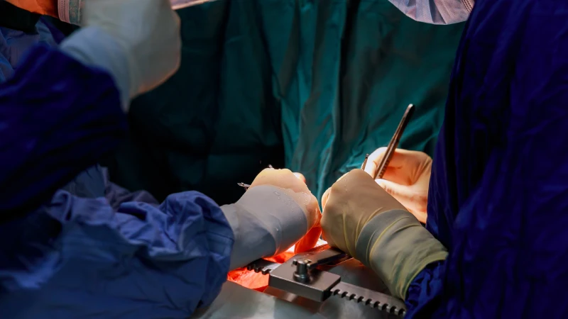
Hirurgija srca
-
Hirurgija srčanih zalistaka
-
Hirurgija aorte
-
Hirurgija karotidnih arterija
-
Hirurgija perifernih arterija
Otvaramo puteve vašem srcu…
Avicenna bolnica je na vašem usluzi sa najnovijom tehnologijom u kardiologiji. Naš tim stručnjaka je ponosan što doprinosi svojim znanjem i veštinama u smanjenju rizika od invazivnih procedura.
KARDIOVASKULARNA HIRURGIJA
Otvaramo puteve vašem srcu… Avicenna bolnica je na vašem usluzi sa najnovijom tehnologijom u kardiologiji. Naš tim stručnjaka je ponosan što doprinosi svojim znanjem i veštinama u smanjenju rizika od invazivnih procedura.
KARDIOLOGIJA
Testovi napora se sprovode u slučajevima sumnje na suženje arterija. Ehokardiografski ultrazvuk i Doppler analiza se sprovode kako bi se utvrdili uslovi srčanih disfunkcija. Poremećaji ritma, povezani sa hipertenzijom, ocenjuju se pomoću Holter monitoringa.
NEINVAZIVNA KARDIOLOŠKA JEDINICA
Dijagnoza i terapija
-
Kardiološka klinika
-
Laboratorija za testove napora
-
Laboratorija za ehokardiografiju, obojena Doppler ehokardiografija, kontrastna i stresna ehokardiografija
Kontrastna i stresna ehokardiografija,
Holter monitori
Holter ritma, Holter krvnog pritiska
Terapijske procedure
-
Koronarna angioplastika
-
Angioplastika – ugradnja stenta
-
Privremeni i trajni pacemaker implantati
-
Koronarna jedinica intenzivne nege
-
Primarni PTCA + stent
-
Pulmonarna balon valvuloplastika
INVAZIVNA KARDIOLOŠKA JEDINICA
Dijagnoza i terapija
-
Koronarna angiografija
-
Srčana kateterizacija
-
Periferna angiografija


Leave a Reply